Children’s Feet
Children with strong, healthy feet often avoid many kinds of lower extremity problems later in life. Contact our office to have your children’s feet and lower extremities examined.
Infants
The size and shape of your baby’s feet change quickly during their first year. Because a baby’s feet are flexible, too much pressure or strain can affect their feet’s shape. It’s important to allow your baby to kick and stretch his or her feet. Also, make sure shoes and socks do not squeeze the toes.
Toddlers
Try not to force your toddler to walk before she is ready. Carefully watch her gait once she begins to walk. If your toddler’s toe touches down instead of the heel, or she always sits while others play, contact our office. Many toddlers have a pigeon-toe gait, and this is normal. Most children outgrow the problem.
When foot care is needed
To help with flatfeet, special shoes or custom-made shoe inserts may be prescribed. To correct mild intoeing, your toddler may need to sit in a different position while playing or watching TV. If you child’s feet turn in or out a lot, corrective shoes, splints, or night braces may be prescribed.
The foot’s bone structure is well-formed by the time your child reaches age 7 or 8, but if a growth plate (the area where bone growth begins) is injured, the damaged plate may cause the bone to grow oddly. With a doctor’s care, however, the risk of future bone problems is reduced.
Remember to check your child’s shoe size often. Make sure there is space between the toes and the end of the shoe, Make sure their shoes are roomy enough to allow the toes to move freely. Don’t let your child wear hand-me-downs.
What Is Calcaneal Apophysitis (Sever’s Disease)?
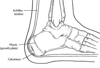
Calcaneal apophysitis is a painful inflammation of the heel’s growth plate. It typically affects children between the ages of 8 and 14 years old, because the heel bone (calcaneus) is not fully developed until at least age 14. Until then, new bone is forming at the growth plate (physis), a weak area located at the back of the heel. When there is too much repetitive stress on the growth plate, inflammation can develop.
Calcaneal apophysitis is also called Sever’s disease, although it is not a true “disease.” It is the most common cause of heel pain in children, and can occur in one or both feet.
Heel pain in children differs from the most common type of heel pain experienced by adults. While heel pain in adults usually subsides after a period of walking, pediatric heel pain generally doesn’t improve in this manner. In fact, walking typically makes the pain worse

Causes
Overuse and stress on the heel bone through participation in sports is a major cause of calcaneal apophysitis. The heel’s growth plate is sensitive to repeated running and pounding on hard surfaces, resulting in muscle strain and inflamed tissue. For this reason, children and adolescents involved in soccer, track, or basketball are especially vulnerable.
Other potential causes of calcaneal apophysitis include obesity, a tight Achilles tendon, and biomechanical problems such as flatfoot or a high-arched foot.
Symptoms
Symptoms of calcaneal apophysitis may include:
Diagnosis
To diagnose the cause of the child’s heel pain and rule out other more serious conditions, the foot and ankle surgeon obtains a thorough medical history and asks questions about recent activities. The surgeon will also examine the child’s foot and leg. X-rays are often used to evaluate the condition. Other advanced imaging studies and laboratory tests may also be ordered.
Treatment
The surgeon may select one or more of the following options to treat calcaneal apophysitis:
- Reduce activity. The child needs to reduce or stop any activity that causes pain.
- Support the heel. Temporary shoe inserts or custom orthotic devices may provide support for the heel.
- Medications. Nonsteroidal anti-inflammatory drugs (NSAIDs), such as ibuprofen, help reduce the pain and inflammation.
- Physical therapy. Stretching or physical therapy modalities are sometimes used to promote healing of the inflamed issue.
- Immobilization. In some severe cases of pediatric heel pain, a cast may be used to promote healing while keeping the foot and ankle totally immobile.
Often heel pain in children returns after it has been treated because the heel bone is still growing. Recurrence of heel pain may be a sign of calcaneal apophysitis, or it may indicate a different problem. If your child has a repeat bout of heel pain, be sure to make an appointment with your foot and ankle surgeon.
Can Calcaneal Apophysitis Be Prevented?
The chances of a child developing heel pain can be reduced by:
- Avoiding obesity
- Choosing well-constructed, supportive shoes that are appropriate for the child’s activity
- Avoiding or limiting wearing of cleated athletic shoes
- Avoiding activity beyond a child’s ability.
Flat Feet (over pronation)
Flat feet are a common condition. In infants and toddlers, the longitudinal arch is not developed and flat feet are normal. Most feet are flexible and an arch appears when the person stands on his or her toes. The arch develops in childhood, and by adulthood most people have developed normal arches.
Most flat feet usually do not cause pain or other problems. Flat feet may be associated with pronation, a leaning inward of the ankle bones toward the center line. Shoes of children who pronate, when placed side by side, will lean toward each other (after they have been worn long enough for the foot position to remodel their shape).
Foot pain, ankle pain or lower leg pain, especially in children, may be a result of flat feet and should be evaluated.
Treatment:
Orthotic devices may be recommended. In severe cases, surgery on the midfoot bones may be necessary to treat the associated flatfoot condition.
Tarsal coalition:
Tarsal coalition is a bone condition that causes decreased motion or absence of motion in one or more of the joints in the foot, usually creating a very flat foot without much motion.
The bones found at the top of the arch, the heel, and the ankle are referred to as the tarsal bones. A tarsal coalition is an abnormal connection between two of the tarsal bones in the back of the foot or the arch. This abnormal connection between two bones is most commonly an inherited trait.
The lack of motion or absence of motion experienced in a tarsal coalition is caused by abnormal bone, cartilage, or fibrous tissue growth across a joint. When excess bone has grown across a joint, it may result in restricted or a complete lack of motion in that joint. Cartilage or fibrous tissue growth can restrict motion of the affected joint to varying degrees, causing pain in the affected joint and/or in surrounding joints.
Symptoms
Treatment:
Nonsurgical treatments, such as corrective shoes or custom orthotics, physical therapy, and anti-inflammatory medication, are the first courses of action. Note: Please consult your physician before taking any medications.
Surgery is sometimes performed in severe cases to allow for more normal motion between the bones or to fuse the affected joint or surrounding joints.
Intoeing:
Intoeing is a condition caused by curving inward of the feet when walking or running.. A professional medical evaluation is recommended if the child walks intoed as well as is”bowed legged”. This condition can be easily treated with special shoes and or braces.
Club foot:
Clubfoot is one of the most common, non-life threatening, major birth defects among infants globally. Approximately one in every 1,000 newborns has clubfoot. Of those, one in three have both feet clubbed. The exact cause is unknown. Two out of three clubfoot babies are boys. Clubfoot is twice as likely to occur if one or both parents and/or a sibling has had it. Less severe infant foot problems are often incorrectly called clubfoot.
Clubfoot twists the heel and toes inward. It often appears like the top of the foot is on the bottom. Additionally, the clubfoot, calf, and leg are smaller and shorter than normal. When clubfoot is detected at birth, it is not painful and is correctable.
The goal of treating clubfoot is to make the infant’s clubfoot (or feet) functional, painless, and stable by the time he or she is ready to walk. Serial casting is the process used to slowly move the bones of a clubfoot into the proper alignment. The doctor starts by gently stretching the child’s clubfoot toward the correct position. A cast is put on to hold the foot in place. One week later, the cast is removed, the baby’s foot is stretched a little farther toward the correct position, and a new cast is applied. X-rays are used throughout the process to check on progress toward proper foot alignment. Casting generally repeats for 6-12 weeks, and may take up to 4 months.
About half the time, clubfoot straightens with casting. Once the proper foot alignment is achieved, the child is fitted with special shoes or braces to keep the foot straight once corrected. These maintenance devices are used until the child has been walking for up to a year or more. Muscles for children with clubfoot commonly try to return to the clubfoot position; a regular occurrence among 2 and 3 year olds, but a condition that may continue up to age 7.
In some cases, stretching, casting, and bracing is not enough to correct clubfoot. Surgery may be required to adjust the tendons, ligaments, and joints in the foot and ankle.
